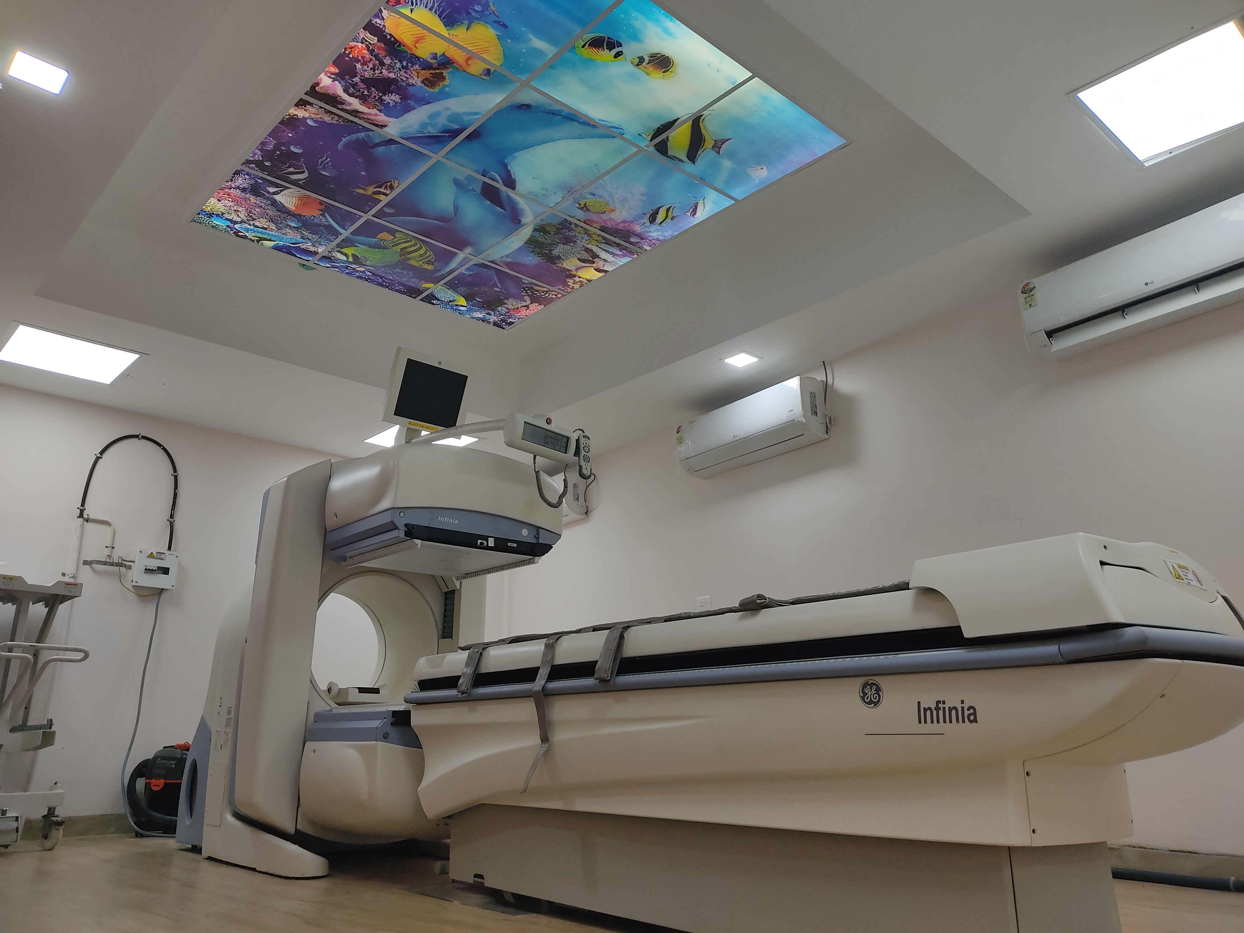
Gamma camera scans are a type of medical imaging procedure that uses a gamma camera to detect gamma rays emitted by a radioactive tracer introduced into the body. These scans are commonly used in nuclear medicine for various diagnostic purposes. Here's an overview of different types of gamma camera scans:
Bone Scan:
- Purpose: Detects abnormalities in bones, such as fractures, infections, tumors, or areas with abnormal bone metabolism.
- Procedure: A small amount of radioactive material is injected intravenously. The gamma camera captures images as the tracer accumulates in bones over time.
Myocardial Perfusion Imaging (MPI) or Cardiac Stress Test:
- Purpose:Assesses blood flow to the heart muscle and identifies areas with reduced blood supply, often used in evaluating coronary artery disease.
- Procedure: The patient undergoes a stress test, either through exercise or medication. A radioactive tracer is injected, and images are taken at rest and during stress to compare blood flow.
Thyroid Scan:
- Purpose: Evaluates the structure and function of the thyroid gland, identifying nodules, inflammation, or overactive or underactive thyroid conditions.
- Procedure: A radioactive iodine tracer is administered orally or intravenously. The gamma camera captures the distribution of the tracer in the thyroid.
Renal (Kidney) Scan:
- Purpose: Assesses kidney function, blood flow, and drainage. Detects conditions such as kidney infections, tumors, or blockages.
- Procedure: A radioactive tracer is injected, and the gamma camera captures images as the tracer is filtered and excreted by the kidneys.
Parathyroid Scan:
- Purpose: Locates overactive parathyroid glands, which may cause hyperparathyroidism.
- Procedure: A small amount of radioactive material is injected, and the gamma camera captures images to identify abnormal parathyroid activity.
Myocardial Perfusion Scan:
This scan is a pivotal tool in assessing blood flow to the heart muscle, aiding in the diagnosis of coronary artery disease. During the procedure, a small amount of a radioactive tracer is injected, and the gamma camera captures detailed images at rest and during stress. B.
Myocardial Viability Scan:
- When heart tissue undergoes damage due to conditions like a heart attack, determining its viability becomes crucial for treatment decisions. The Myocardial Viability Scan is designed for precisely that purpose. Through the use of a radioactive tracer, our gamma camera captures images that reveal viable and non-viable myocardial tissue. This information is instrumental in guiding therapeutic strategies, ensuring optimal care for patients with compromised heart function.
Gamma camera scans are valuable diagnostic tools, providing functional and anatomical information about various organs and tissues. These scans play a crucial role in personalized medicine, helping healthcare professionals tailor treatment plans based on individual patient conditions.
Feel free to contact us!
NUCLEUZ HEALTHCARE PRIVATE LIMITED,TC 17/127(1&2), Ulloor Junction, Near Credence Hospital, Opposite Catholic Syrian Bank, Medical College P.O,
Thiruvananthapuram, Kerala 695011.